Listening to the Invisible: Wave Propagation, Interference, and the Science That Unites Sonar, Ultrasound, and Acoustic Engineering
Light is the dominant sense modality through which humans engage with the physical world, and it is correspondingly the medium most associated with scientific instrumentation. Yet light is a poor probe of many environments that matter enormously to medicine, national defense, and industrial engineering. Seawater attenuates electromagnetic radiation rapidly; human tissue scatters optical wavelengths over distances of millimeters; the interior of a load-bearing weld is opaque to visual inspection. In each of these domains, acoustic waves—longitudinal pressure oscillations propagating through a material medium—perform tasks that no photon can. Understanding why requires engaging seriously with the physics of wave behavior, from the basic mechanics of propagation to the subtleties of interference and frequency-dependent attenuation.
The Mechanics of a Pressure Wave
A sound wave is a disturbance in which particles of a medium are alternately compressed and rarefied along the direction of propagation. Unlike electromagnetic waves, which require no medium and travel at the same speed in vacuum regardless of frequency, acoustic waves are entirely dependent on the mechanical properties of their host material. The speed of sound in a medium is determined by the ratio of the medium's bulk modulus—its resistance to compression—to its density. In dry air at 20°C, this yields approximately 343 meters per second. In water, the much higher bulk modulus produces a propagation speed near 1,480 meters per second. In steel, the speed exceeds 5,000 meters per second.
This dependence on material properties is not merely a curiosity; it is the physical basis for nearly every acoustic sensing technology. When a pressure wave encounters an interface between two media with different acoustic impedances—defined as the product of density and wave speed—a fraction of the wave's energy reflects and a fraction transmits. The reflection coefficient is determined by the impedance mismatch: a large mismatch produces a strong reflection, a small mismatch allows most energy to pass. This is why ultrasonic gel is applied to the skin before a medical scan—it eliminates the air gap between the transducer and tissue, reducing an otherwise prohibitive impedance mismatch that would reflect nearly all incident energy before it could enter the body.
Sonar: Acoustic Ranging in a Hostile Medium
The development of sonar—Sound Navigation and Ranging—emerged from a military imperative that became acute during World War I, when German U-boats threatened Allied shipping in the Atlantic. The fundamental operating principle of active sonar is straightforward: a transducer emits an acoustic pulse, and the time elapsed before a reflected echo returns encodes the distance to the reflecting object, given the known propagation speed. Range equals half the product of propagation speed and round-trip travel time.
In practice, however, the ocean is a far more complex acoustic environment than this simple model implies. Sound speed in seawater varies with temperature, salinity, and pressure, all of which change with depth. These variations refract acoustic rays—bending them toward regions of lower propagation speed—in a manner analogous to the refraction of light at an optical interface. The result is the formation of acoustic channels, most notably the SOFAR channel (Sound Fixing and Ranging), located at the depth where sound speed reaches a minimum, typically between 600 and 1,200 meters in the open ocean. Sound trapped within this channel can propagate thousands of kilometers with relatively little attenuation, a phenomenon exploited both by military long-range detection systems and by marine biologists studying whale communication.
Passive sonar, by contrast, emits nothing. It listens for acoustic signatures produced by a target—the cavitation noise of a propeller, the mechanical vibration of machinery, the flow noise of water moving over a hull. Passive detection preserves the observer's own acoustic stealth, which is why it is the preferred mode for submarines operating in contested waters. Signal processing algorithms, including fast Fourier transform (FFT) analysis of the received signal's frequency content, allow operators to identify characteristic spectral signatures that distinguish one vessel class from another.
Medical Ultrasound: Imaging Through Tissue
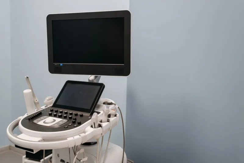
Medical ultrasound repurposes the same reflection physics for diagnostic imaging, operating at frequencies between 1 and 20 megahertz—far above the audible range, which extends to approximately 20 kilohertz in young adults. The choice of frequency involves a fundamental trade-off rooted in wave physics: spatial resolution scales with wavelength, which decreases as frequency increases, but attenuation in tissue also increases with frequency. A 15 MHz transducer resolves structures approximately 0.1 millimeters in size but cannot image more than a few centimeters deep; a 2 MHz transducer reaches 20 centimeters or more but sacrifices resolution proportionally. Clinical sonographers select frequency based on the depth of the target anatomy.
Modern phased-array ultrasound systems address a further challenge: producing a two-dimensional image from a one-dimensional array of transducer elements. By applying precisely calculated time delays to the electrical pulses driving individual array elements, the system steers and focuses the emitted acoustic beam electronically, without mechanical movement. The same delay scheme applied to the received signals allows the system to synthesize echoes from a specific focal depth, suppressing out-of-focus contributions. This beamforming process is an exercise in constructive and destructive interference: signals from the desired direction add coherently, while those from other angles cancel.
Doppler ultrasound extends this capability by measuring the frequency shift of echoes returned from moving structures—most commonly red blood cells flowing through vessels. The Doppler effect, familiar from the pitch change of a passing siren, shifts the reflected frequency by an amount proportional to the reflector's velocity component along the beam axis. Color flow Doppler maps these velocity data onto the anatomical image, allowing clinicians to visualize blood flow direction and speed in real time—a capability with obvious diagnostic value in cardiology, vascular surgery, and obstetrics.
Industrial Acoustics: Flaw Detection and Structural Monitoring
Non-destructive testing (NDT) using ultrasound applies the same reflection principles to industrial quality control. A transducer coupled to the surface of a metal component emits a pulse that propagates through the bulk material. Internal discontinuities—cracks, voids, inclusions, or delaminations—reflect energy back to the transducer. The amplitude and timing of these echoes reveal the size and depth of the flaw without damaging the part under inspection.
This technique is mandated for critical components in aerospace, nuclear power, and pressure vessel manufacturing. Phased-array ultrasonic testing (PAUT) applies the same electronic steering used in medical imaging to industrial inspection, producing cross-sectional views of welds and forgings that would otherwise require destructive sectioning to examine. The Federal Aviation Administration and the Nuclear Regulatory Commission both rely on ultrasonic inspection data as part of their safety certification frameworks.
Acoustic emission monitoring takes a complementary approach: rather than actively interrogating a structure, it passively listens for the stress waves emitted when a crack propagates or a material yields. Sensors distributed across a structure triangulate the source location from the differential arrival times of the emitted wave—a method directly analogous to seismological location of earthquake epicenters.
Interference, Diffraction, and the Limits of Resolution
All acoustic imaging systems confront a resolution limit imposed by diffraction. A wave cannot resolve features smaller than roughly half its wavelength—a constraint known as the Rayleigh criterion in optics but equally applicable to acoustics. This limit is not a failure of engineering but a consequence of wave physics: diffraction causes a finite-aperture beam to spread, and two closely spaced reflectors whose separation falls below the diffraction limit produce overlapping echo patterns that cannot be distinguished.
Researchers have developed techniques to partially circumvent this limit. Synthetic aperture sonar and ultrasound reconstruct a large effective aperture from a moving transducer, improving lateral resolution beyond what any physically realizable aperture could achieve at a given frequency. Acoustic metamaterials—engineered structures with subwavelength features that manipulate wave propagation in ways natural materials cannot—have demonstrated the ability to focus sound below the diffraction limit in controlled laboratory settings, opening potential pathways to super-resolution acoustic imaging.
The physics of sound, in this sense, is not a closed subject. It is a domain in which fundamental wave mechanics continues to generate both new applications and new questions.